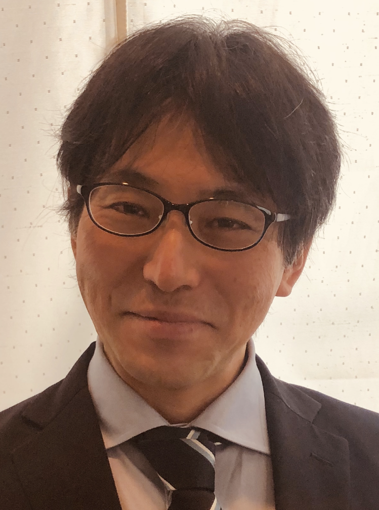
Prof. Daisuke Matsubara
Department of Diagnostic Pathology,
Faculty of Medicine, University of Tsukuba
In recent years, genetic testing and immunohistochemistory have become indispensable for treatment selection, and pathologists are required to respond to such requests, but the scientific basis for such requests is often limited to the fact that they have been approved by the FDA or that they have been determined to be so in clinical trials. As a result of this passive attitude, pathologists are losing the habit of thinking for themselves based on genomic and clinical information, conference presentations, and articles. Integrated classification and integrated understanding of genomics, clinical practice, and pathology (morphology and phenotype) is a well-known phrase, but data science is becoming the sole domain of pathologists. However, the information on pathology (morphology and phenotype), which is the source of data, is limited to histological types and differentiation levels at best. Can it really be called integrated? Pathologists should communicate more. There are things that only we, pathologists, can understand. We are familiar with the heterogeneity of cancer and can analyze background lesions, microscopic lesions, early stage lesions, and the process of progression in individual cases and in many cases. This kind of realistic analysis based on pathological observations and accumulated experience is important and something to be proud of. On top of this, we would like to incorporate the latest technology and conduct pathological research unique to Japan. For example, TCGA data from overseas is mostly analysis of advanced cancers, with very few cases of early-stage cancers. In Japan, where there are many early-stage cancers, it should be possible to conduct unique Japanese research focusing on early-stage lesions and microdefects while utilizing technologies such as whole genome sequencing analysis and spatial genomics/transcriptomics. EVG staining is useful in identifying invasion, which is well known to all pathologists in Japan. EVG staining is useful for identifying invasion. Taking advantage of this fact, if we collect a large number of EVG-stained specimens and analyze them by AI, we may be able to construct an AI diagnosis system that can make a better diagnosis than one based simply on HE specimens (this is already being promoted in our laboratory). It is also a specialty of Japanese pathologists to value individual cases and pursue them in depth.
Even though pathologists are busy, it is important to collect information, and it is important to keep one's so-called antenna up. The importance of morphological observation by pathologists will never be lost if they maintain a fresh interest, curiosity, and sensitivity to new information.
Profile
Education
2000 MD, Graduate school of Medicine, The University of Tokyo, Japan
Professional experiences
2012 Devision of Molecular Pathology, The Institute of medical science, The University of Tokyo, associate professor
2012 Core laboratory I of pathology, The Institute of medical science, The University of Tokyo, chief
2015 Devision of Integrative Pathology, Department of pathology, Jichi medical University, associate professor
2015 Devision of Molecular Pathology, The Institute of medical science, The University of Tokyo, visiting associate professor
2021 Devision of Diagnostic pathology, Faculty of Medicine, University of Tsukuba, Professor
2021 Devision of Integrative Pathology, Department of pathology, Jichi medical University, associate professor, visiting Professor
Selected publications
- Matsubara, Daisuke; Yoshimoto, Taichiro; Soda, Manabu; Amano, Yusuke; Kihara, Atsushi; Funaki, Toko; Ito, Takeshi; Sakuma, Yuji; Shibano, Tomoki; Endo, Shunsuke; Hagiwara, Koichi; Ishikawa, Shumpei; Fukayama, Masashi; Murakami, Yoshinori; Mano, Hiroyuki; Niki, ToshiroReciprocal expression of TFF-1 (trefoil factor-1) and TTF-1 (thyroid transcription factor-1) in lung adenocarcinomas. Canser Sci, 2020 in press (IF:4.966)
- Takeshi Ito, Atsuko Nakamura, Ichidai Tanaka, Yumi Tsuboi, Teppei Morikawa, Jun Nakajima, Daiya Takai, Masashi Fukayama, Yoshitaka Sekido, Toshiro Niki, Daisuke Matsubara*, and Yoshinori Murakami. CADM1 associates with Hippo pathway core kinases; membranous co-expression of CADM1 & LATS2 in lung tumors predicts good prognosis. Canser Sci, 2019;110:2284-2295. doi: 10.1111/cas.14040. (corresponding author, co-last author) Cancer Science2019年7月号のハイライト論文(IF:4.966)
- Matsubara D, Soda M, Yoshimoto T, Amano Y, Sakuma Y, Yamato A, Ueno T, Kojima S, Shibano T, Hosono Y, Kawazu M, Yamashita Y, Endo S, Hagiwara K, Fukayama M, Takahashi T, Mano H, Niki T. Inactivating mutations and hypermethylation of the NKX2-1/TTF-1 gene in non-TRU-type lung adenocarcinomas. Cancer Sci, 2017 Jul 5. doi: 10.1111/cas.13313. (IF:4.966)
- Ito T, Matsubara D*, Tanaka I, Makiya K, Tanei ZI, Kumagai Y, Shiu SJ, Nakaoka HJ, Ishikawa S, Isagawa T, Morikawa T, Shinozaki-Ushiku A, Goto Y, Nakano T, Tsuchiya T, Tsubochi H, Komura D, Aburatani H, Dobashi Y, Nakajima J, Endo S, Fukayama M, Sekido Y, Niki T, Murakami Y. Loss of YAP1 Defines Neuroendocrine Differentiation of Lung Tumors. Cancer Sci, 2016;107:1527-1538. doi: 10.1111/cas.13013. (corresponding author) (IF:4.966)
- Matsubara D, Kishaba Y, Yoshimoto T, Sakuma Y, Sakatani T, Tamura T, Endo S, Sugiyama Y, Murakami Y, Niki T. Immunohistochemical analysis of the expression of E-cadherin and ZEB1 in non-small cell lung cancer. Pathol Int. 64: 560-8, 2014. (IF:2.110)
- Ibrahim R, Matsubara D*, Osman Y, Morikawa T, Goto A, Morita S, Ishikawa S, Aburatani H, Takai D, Nakajima J, Fukayama M, Niki T, Murakami Y. Expression of PRMT5 in lung adenocarcinoma and its significance in epithelial-mesenchymal transition. Human Pathol. 45: 1397-1405, 2014 (corresponding author) (IF:2.740)
- Matsubara D, Kishaba Y, Ishikawa S, Sakatani T, Oguni S, Tamura T, Hoshino H, Sugiyama Y, Endo S, Murakami Y, Aburatani H, Fukayama M, Niki T. Lung Cancer with Loss of BRG1/BRM, shows Epithelial Mesenchymal Transition Phenotype and Distinct Histologic and Genetic Features. Cancer Sci. 2013;104(2):266-73. (IF:4.966)
- Matsubara D, Kanai Y, Ishikawa S,Ohara S, Yoshimoto T, Sakatani T, Oguni S,Tamura T, Kataoka H, Endo S, Murakami Y, Aburatani H, Fukayama M and Niki T. Identification of CCDC6-RET Fusion in a Human Lung Adenocarcinoma Cell Line, LC-2/ad.J Thorac Oncol. 2012;7(12):1872-6. (IF:13.357)
- Matsubara D, Niki T. Epidermal Growth Factor Receptor Mutation and Chemosensitivity. J Thorac Oncol. 2012;7(4):771-772. (IF:13.357)
- Matsubara D, Ishikawa S, Oguni S, Aburatani H, Fukayama M, Niki T. Co-activation of epidermal growth factor receptor and c-MET defines a distinct subset of lung adenocarcinomas. Am J Pathol. 2010;177(5):2191-2204. (IF:3.762)
- Matsubara D, Ishikawa S, Oguni S, Aburatani H, Fukayama M, Niki T. Molecular predictors of sensitivity to the MET inhibitor PHA665752 in lung carcinoma cells. J Thorac Oncol. 2010;5:1317-24.Journal of thoracic oncoly 2010年9月号のハイライト論文(IF:13.357)
- Matsubara D, Morikawa T, Goto A, Nakajima J, Fukayama M, Niki T. Subepithelial myofibroblast in lung adenocarcinoma: a histologic indicator of excellent prognosis. Mod Pathol. 2009;22:776-85. (IF:6.365)
- Matsubara D, Niki T, Ishikawa S, Goto A, Ohara E, Yokomizo T, Heizmann CW, Aburatani H, Moriyama S, Moriyama H, Nishimura Y, Funata N, Fukayama M Differential expression of S100A2 and S100A4 in lung adenocarcinomas: clinicopathologic significance, relationship to p53, and identification of their target genes. Cancer Sci. 2005;96:844-57. (IF:4.966)
